Today, over half a million women die of breast cancer every year. Yet, if a tumor is less than 1cm in size when it is detected, with no lymph involvement, survival rates at 5 years are comparable with someone who has not had cancer. Micrima believes the way to reduce this number of deaths is frequent screening from an early age. To that end, they have developed a system for breast imaging using harmless radio-waves.
Micrima believes that to provide early detection of breast cancer you need to be able to offer comfortable, frequent screening from an early age. Interestingly in one report 46% of women who did not return for a second round of screening sited pain as the reason. With all this in mind, Micrima has set out to develop a system that is comfortable for the patient, easy to interpret, good in dense tissue and can be used frequently from a much younger age in order to detect cancer early.
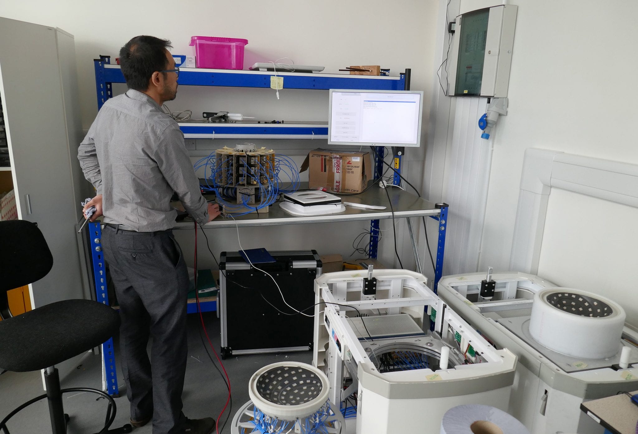
Originally based on technology for the detection of buried landmines, the early application to breast imaging was spun-out from the University of Bristol, as Micrima. The founders of the company recognized the impact the technology could potentially have on breast cancer detection and mortality rates. When the company was spun out of Bristol university the decision was made to stay in Bristol as this was where the relevant engineering talent was. Today, the company consists of 23 hardware, software, and product development engineers with a small commercial team. It is continuing to expand in all areas of the business to meet the strategic aims of the company.
MARIA®: A UNIQUE PRODUCT
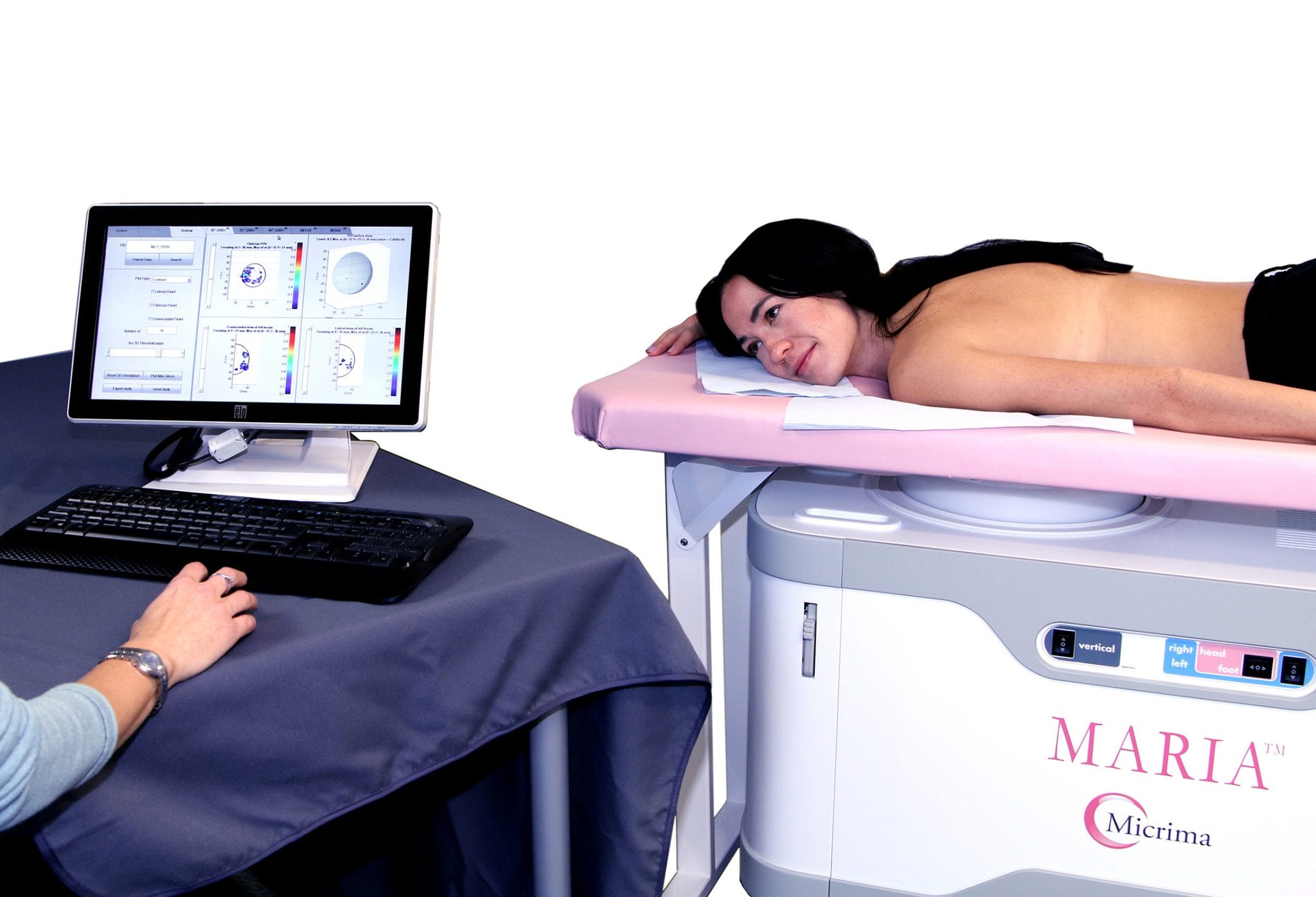
MARIA® (Multistatic Array Radio-wave Image Acquisition) is a revolutionary product from Micrima that uses radio-waves to detect breast cancer. The data acquisition requires the patient to lie on their tummy on the bed, with the breast that is to be imaged pendulant through a hole in the bed. The breast is cupped by a ceramic insert, underneath which there is a radio-wave array containing 60 antennae that each transmits and receives. They each do this in sequence, taking 22 seconds before the array rotates. The same process is repeated twice more to build up a 3D map of the tissues throughout the breast in one min six seconds. MARIA® measures the signal reflected at the interface between tissues of different electromagnetic material properties showing reflections from the edges of lesions. This data is currently shown as an image but there is a wealth of information not currently displayed. The next step in the development of this product is, working closely with clinical partners, to confirm what other data will enhance the cancer detection and improve the interpretation time.
Today the MARIA® breast scanning system needs to be used in conjunction with other imaging methods for cancer detection but it is hoped through further data collection and development of the algorithms the system can start to identify different structures in the breast and begin to identify the likelihood of a structure being malignant or not. Then it can be considered as a stand-alone modality and eventually a screening tool.
Professor Lain Lyburn, a consultant radiologist at Thirlestaine Breast Centre, Cheltenham, recently said, “We have been involved in the evaluation of the MARIA® system for some time now and whilst it is currently offered as an adjunct to other imaging modalities, particularly where dense tissue is involved, the technique promises the exciting ability to distinguish between tissue types in the future. Any imaging modality that can readily give this sort of functional information has the potential to influence many points in the diagnostic and treatment pathways – there could be less need for biopsies and possibly a reduction in overtreatment. ”
In recent years, Micrima has been recognized with several awards. Most of these awards are focused on the technology and innovation of the MARIA® system. These include general innovation awards, like the ‘Medilink SW innovation Award in 2018’. Micrima has also been honored with ‘The Frost and Sullivan New Product Innovation Award- Breast Imaging in 2019’ and the ‘Global Health and Pharma Best Oncology Diagnostic Imaging Device Manufacture in 2019 Award’.
The ongoing primary goal for Micrima is the evolution of the MARIA® software to deliver increasing degrees of tissue differentiation. The company is hopeful to be able to identify the type of malignancy along with the diagnosis of the disease. “If we can assist radiologists by indicating which are low grade, slow-growing, and can/should be monitored over time, and which are invasive, high grade, and rapidly growing, this could have huge implications for the treatment of breast cancer. There is also the possibility to use this technology for detecting other types of cancer in the body in the future,” says Roy Johnson Micrima’s Executive Chairman.